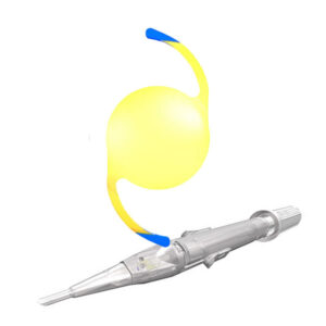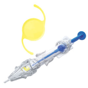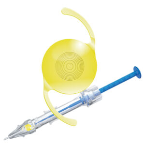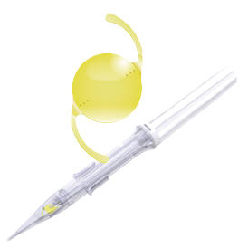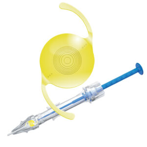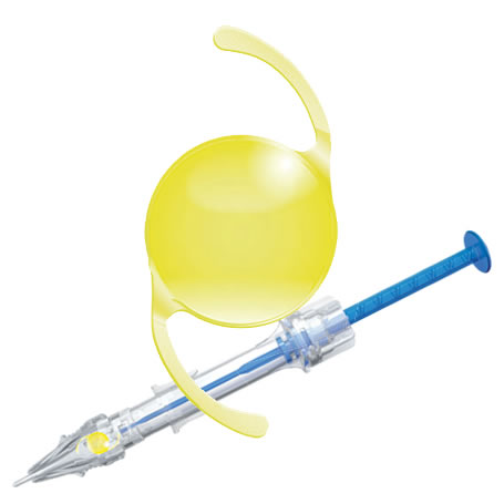
LIO monofocal enhanced implantada com o injetor preloaded multiSert™
- Vivinex Impress™ proporciona excelente visão intermediária aos pacientes monofocais
- Vivinex Impress™ proporciona uma melhora na acuidade visual binocular a 66 cm
- Vivinex Impress™ proporciona a mesma acuidade visual corrigida para longe que uma LIO asférica monofocal padrão1
- Vivinex Impress™ melhora a acuidade visual intermediária a 66 cm (-1,5 D de defocus) em mais de 1 linha1
- Vivinex Impress™ melhora a visão intermediária e proporciona previsibilidade refrativa
Benefícios da plataforma Vivinex™:
Glistening-free: Material da LIO em acrílico hidrofóbico livre de glistening.3, 4
Melhor qualidade de Imagem: O desenho óptico asférico patenteado da Vivinex™ compensa parcialmente a aberração esférica da córnea e é mais tolerante as fontes de coma do que os desenhos asféricos padrões.5
Redução do PCO: Tratamento com oxigênio ativo, uma superfície lisa e uma borda óptica quadrada para reduzir a opacidade de cápsula posterior – PCO.3,6,7,8,9,10,11,12
Abertura suave da LIO e estabilidade no saco capsular: Superfície da alça texturizada e rugosa para
reduzir o potencial de adesão à superfície óptica durante o implante e proporcionar melhor aderência ao saco capsular.
Implante com o injetor preloaded multiSertTM
Modos empurrar e rosquear com controle de profundidade de inserção.
Vivinex™ multiSert™ é um sistema 4 em 1 que permite atingir excelente consistência no implante com escolha no estilo de implante e inserção13
Os injetores preloaded são:
Mais fáceis de preparar, aumentando a segurança com:14,15,16,17,18,19
• Redução do risco de infecção e contaminação
• Redução do risco de danos à LIO
Mais eficientes na sala de cirurgia:16,18
• Minimizando o tempo necessário para preparar o sistema de implante da LIO
• Exigindo menos instrumentos que precisam ser reprocessados
Mais previsíveis:18
• Aumentando a previsibilidade e consistência no implante da LIO
Para mais informações visite: https://adaptltda.com.br/impress/
Referência:
- HOYA data on file. CTM-23-P0105, HOYA Medical Singapore, Pte. Ltd, 2023
- HOYA data on file RnD-20-367, HOYA Medical Singapore, Pte. Ltd, 2023
- Tandogan, T. et al. (2021): In-vitro glistening formation in six different foldable hydrophobic intraocular lenses. In BMC Ophthalmol 21, 126.
- Auffarth et al. (2023) Randomized multicenter trial to assess posterior capsule pacification and glistenings in two hydrophobic acrylic intraocular lenses. Sci Rep 13, 2822.
- Pérez-Merino, P.; Marcos, S. (2018): Effect of intraocular lens decentration on image quality tested in a custom model eye. In: Journal of cataract and refractive surgery 44 (7), p. 889-896.
- Leydolt, C. et al. (2020): Posterior capsule pacification with two hydrophobic acrylic intraocular lenses: 3-year results of a randomized trial. In: American journal of ophthalmology 217 (9), p. 224-231.
- Giacinto, C. et al. (2019): Surface properties of commercially available hydrophobic acrylic intraocular lenses: Comparative study. In: Journal of cataract and refractive surgery 45 (9), p. 1330-1334.
- Werner, L. et al. (2019): Evaluation of clarity characteristics in a new hydrophobic acrylic IOL in comparison to commercially available IOLs. In: Journal of cataract and refractive surgery 45 (10), p. 1490-1497.
- Matsushima, H. et al. (2006): Active oxygen processing for acrylic intraocular lenses to prevent
posterior capsule pacification. In: Journal of cataract and refractive surgery 32 (6), p. 1035-1040. - Farukhi, A. et al. (2015): Evaluation of uveal and capsule biocompatibility of a single-piece
hydrophobic acrylic intraocular lens with ultraviolet-ozone treatment on the posterior surface. In: Journal of cataract and refractive surgery 41 (5), p. 1081-1087. - Eldred, J. et al. (2019): An In Vitro Human Lens Capsular Bag Model Adopting a Graded Culture Regime to Assess Putative Impact of IOLs on PCO Formation. In: Investigative ophthalmology & visual science 60 (1), p. 113-122.
- Nanavaty, M. et al. (2019): Edge profile of commercially available square-edged
intraocular lenses: Part 2. In: Journal of cataract and refractive surgery 45 (6), p. 847-853. - HOYA data on file. DoF-SERT-102-MULT-03052018, HOYA Medical Singapore Pte. Ltd, 2018
- Galor, A. er al. (2013). Management strategies to reduce risk of postoperative infections. In Current ophthalmology reports, 1(4), 10.1007/40135-013-0021-5.
- Bodnar, Z. et al. (2012). Toxic anterior segment syndrome: Update on the most common causes. In: Journal of cataract and refractive surgery, 38(11), 1902-1910.
- Jones, J. et al. (2016). The impact of a preloaded intraocular lens delivery system on operating room efficiency in routine cataract surgery. In: Clinical ophthalmology (Auckland, N.Z.), 10, 1123-1129.
- Park, C. et al. (2018). Toxic anterior segment syndrome-an updated review. In: BMC ophthalmology, 18(1), 276.
- Chung, B. et al. (2018). Preloaded and non-preloaded intraocular lens delivery system and characteristics: human and porcine eyes trial. In: International journal of ophthalmology, 11(1), 6-11.
- Schmidbauer, J. et al. Rates and causes of intraoperative removal of foldable and rigid intraocular lenses: clinicopathological analysis of 100 cases. In: Journal of cataract and refractive surgery, 28(7), 1223-1228.