Nós temos em torno de 15 anos de estudos publicados sobre a consistente tecnologia da Diopsys, com aceitação e uso histórico da eletroretinografia (ERG) e potencial visual evocado (VEP). Somados a, diversos artigos e pesquisas sendo publicadas anualmente, encaminhando instituições de cuidados oftalmológicos a apoiarem os mais diversos benefícios que a eletrofisiologia visual poderia proporcionar aos oftalmologistas.


Richard Tipperman, MD | Inside ffERG: Quantify, Predict and Treat Retinal Dysfunction
Ophthalmologist Dr. Rich Tipperman from Wills Eye Hospital reviews how electrophysiology testing in the ophthalmic practice fulfills a diagnostic need that no other device provides. He focuses on quantifying disease and tracking treatment.

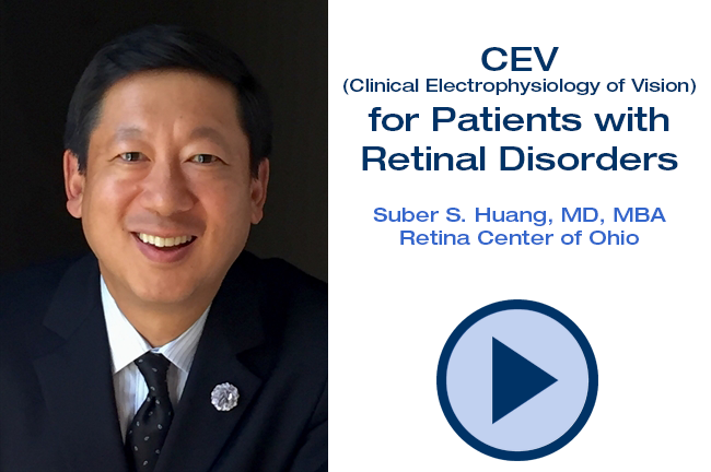
Suber Huang, MD, MBA | CEV for Patients with Retinal Disorders
Watch as retina specialist Dr. Suber Huang provides an overview of the importance of visual electrophysiology testing in the ophthalmic practice, focusing on the need for objective, functional metrics for patients with retinal disorders.

Multi-Luminance Flicker Electroretinography (ERG) Evaluates Impact of anti-VEGF Injections
Diopsys® ffERG quantifies retinal function improvement in diabetic retinopathy. Patient Profile A 67-year-old male patient made an appointment with a chief complaint of blurry vision OU. He had a past medical history of diabetes (12 years) and systemic hypertension (7 years). Past ocular history consisted of moderate diabetic retinopathy (5 years) and dry eyes. Examination
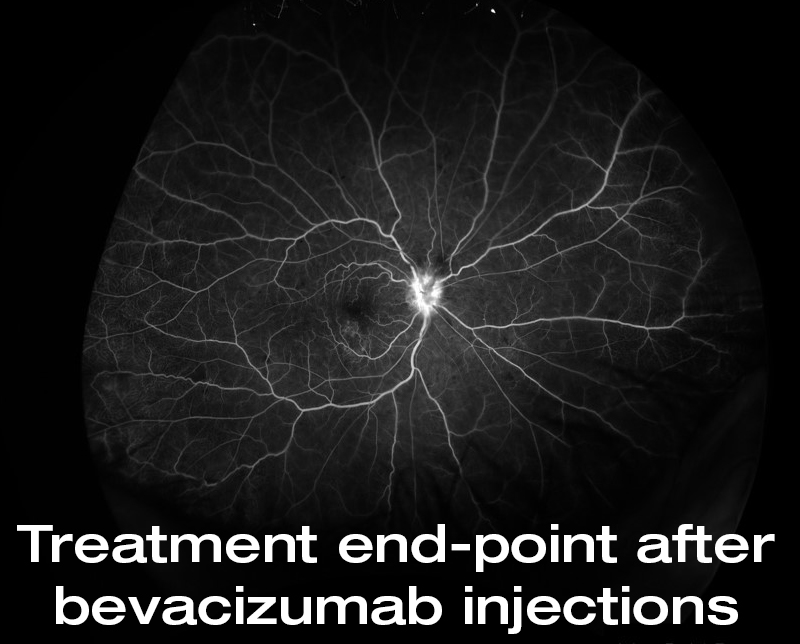
Full Field Electroretinography (ffERG) Verifies Treatment Success in CRVO
Diopsys® ffERG shows treatment end-point when functional improvements stabilize after multiple Avastin® (bevacizumab) injections. Patient Profile An 88-year-old male patient presented with a chief complaint of blurred central vision OD for 1-2 weeks. He had a past medical history of coronary artery disease. Past ocular history consisted of S/P cataract extraction OU, laser posterior capsulotomy

Electroretinography: Out of the laboratory and into the clinic
Ophthalmologist William Ayliffe, FRCS, PHD discusses the widespread uses of electroretinography (ERG) in clinical practice including glaucoma, central retinal vein occlusion, retinal ischaemia, and macular degeneration. He notes that his advanced device (Diopsys NOVA) has allowed him to use this testing in his clinic daily. “These tests are unique in their ability to not only measure disease progression, but also improvement of cellular function.”
A Flicker of Light on Retinal Function
Mitchell Jackson, MD discusses how full-field flicker electroretinography (ffERG) allows him to evaluate patients’ retinal health even in the presence of a dense media opacity cataract. He writes that the ffERG results help him in two ways. “First, they enable me to set realistic expectations for my patients. If their retinas are healthy, cataract surgery
Evaluating the Retina Before Cataract Surgery
Most high-volume cataract surgeons will encounter cataracts that limit or prohibit the view of the fundus fairly regularly. The risk-benefit analysis in these eyes is greatly facilitated by the availability of objective, quantifiable data about the function of the retina, particularly the cone cells. In my clinic, fERG is a routine part of evaluating such cases for planning surgery and managing expectations.
MEDBLOG: Advancements in devices that promote diagnosis and management of retinal disorders
Robert Noecker, MD, MBA, focuses his blog on the positive impact diagnostics and imaging have on various disease states and conditions. In this article, he discusses the use of OCT, pattern electroretinography (PERG), visual evoked potential (VEP), and corneal hysteresis as technologies that enhance the ophthalmologist’s ability to more accurately diagnose and treat patients, while
Role of Electrophysiology in the Early Diagnosis and Follow-Up of Diabetic Retinopathy
“Electroretinography (ERG) and visual evoked potentials (VEP) allow a more detailed study of the visual function and of the possible effects that diabetes can have on the visual function. These techniques have an important role both in the clinic and in research: the central nervous system, in fact, has received much less attention than the peripheral one in the study of the complications of diabetes.These techniques are safe, repeatable, quick, and objective. In addition, both the ERG (especially the oscillatory potentials and the flicker-ERG) and VEP have proved to be successful tools for the early diagnosis of the disease and, potentially, for the ophthalmologic follow-up of diabetic patients.”
N Pescosolido, A Barbato, A Stefanucci, G Buomprisco, Journal of Diabetes Research
MEDBLOG: Improving electrophysiology testing for patients and doctors
Decades ago, electrophysiological testing was only conducted in research facilities and large medical centers, making them very inconvenient for all involved. However, in recent years the testing experience for doctors and patients alike has been vastly improved as VEP, PERG, ffERG, and mfERG are now available in an office setting with the Diopsys NOVA ERG
Electrophysiological Testing of Eyes with Opaque Media
Electrodiagnostic tests of visual function are of undoubted value when applied to eyes with opaque media and suspected retinal disease. The combined investigation of the flash ERG and the YEP has been shown to be of real practical value.
Galloway, N.R. (1988). Electrophysiological testing of eyes with opaque media. Eye, 2, 615-624.
Electroretinographic Evaluation in Adult Diabetics
The electroretinographic component that demonstrates retinal dysfunction in the earlier stage may be a valuable indicator. In the early stage, a delay in the a-wave time and a reduction in the second oscillatory potential amplitude were the most frequent abnormalities: analysis of variance demonstrated that the summed amplitude of the oscillatory potentials and second oscillatory potential amplitude and time were the most sensitive measures of the diabetic retina. Hence, the second oscillatory potential amplitude may be the most sensitive and valuable indicator representing a quantitative measure of overall retinal dysfunction
Kim, SH., Lee, SH., Bae, JY. et al. Doc Ophthalmol (1997) 94: 201. https://doi.org/10.1007/BF02582979
Peripheral Retinal Function Assessed with 30-Hz flicker seems to improve after treatment with Lucentis in Patients with Diabetic Macular oedema
Diabetic retinopathy (DR), especially diabetic macular oedema, is the leading cause of visual loss in the adult population in the Western world.
Holm, K., Schroeder, M. & Lövestam Adrian, M. Doc Ophthalmol (2015) 131: 43. https://doi.org/10.1007/s10633-015-9495-9
Temporal Aspects of Electroretinogram in Diabetic Retinopathy
The correlation of 30-Hz flicker implicit times with retinopathy severity were significant for retinopathy level graded in color fundus photographs as well as for retinal capillary nonperfusion and leakage graded in fluorescein angiograms. A comparison of 30-Hz flicker implicit times in 15 patients with one eye treated with panretinal laser photocoagulation and the other eye untreated (treatment was randomly assigned) showed a significant delay in the treated eyes compared with the untreated eyes (paired eye comparison). This suggests that panretinal laser photocoagulation induces a further delay in the b-wave implicit times of eye treated for diabetic retinopathy.
Bresnick GH, Palta M. Temporal Aspects of the Electroretinogram in Diabetic Retinopathy. Arch Ophthalmol. 1987;105(5):660–664. doi:10.1001/archopht.1987.01060050078042
In So Many Words – Interview with Diopsys’ Joseph Fontanetta
As president and CEO of Diopsys since 2000, Joseph Fontanetta has strived to change how ophthalmologists view their patients. With the company’s ARGOS and NOVA vision testing systems, Mr. Fontanetta and the Diopsys team have brought objective retinal testing from the hospital to the clinic.

How pattern, flicker ERG can impact cataract treatment decisions
William Bond, MD highlights the benefits of pattern and flicker ERG to the cataract surgeon in his latest article. Dr. Bond talks about the basics of ERG, why you would use PERG vs. flicker, and how he chooses patients for each test. He concludes, “I have found the flicker ERG in particular to be reliable
MEDBLOG: Value of office-based ERG and VEP
In this blog article, Robert Noecker, MD, MBA focuses on the accessibility of visual electrophysiology tests, and the benefits to practices with diabetic, cataract, and glaucoma populations. Dr. Noecker states, ERGs and VEPs “are an integral part of being a state-of-the-art facility and key for physicians who want to provide true value to their patients.”
If You Build It, They Will Come
A commitment to excellent outcomes fuels Palm Beach’s Goldman Eye, and using the best equipment helps achieve those outcomes. David Goldman, MD’s advice to colleagues: “go slow and maximize the potential for low overhead for as long as possible; and invest in the best technology.’’ Read the full practice profile from Cataract & Refractive Surgery
Integrate VEP and PERG
In this article by Nathan Lighthizer, O.D., F.A.A.O., he says, “Today, visually evoked potentials (VEP) and pattern electroretinography (PERG) provide excellent diagnostics, are patient friendly, affordable and integrable in the office setting. What’s more, the technology is incredibly useful for glaucoma suspects and those who show signs of early dysfunction because of AMD and diabetic
Adding electroretinography: Technology to clinical utility
Advancements in electrophysiological technology can help ophthalmologists to discover vision-related diseases in their earliest stages. In this article, Blake K. Williamson, MD, shares how use of a vision testing system has made a difference in his practice as a cataract specialist, whereas William E. Sponsel, MD, shares his perspective as a glaucoma specialist.
MEDBLOG: Evidence-based medicine: Increasing value through early diagnostics
Robert Noecker, MD, MBA, focuses his blog on the positive impact diagnostics and imaging have on various disease states and conditions. In this article, he writes about three diagnostic tests he recently started using that have made the most significant difference in helping him identify at-risk patients and determining treatment therapies for early glaucoma: measuring
Clinical: Glaucoma – Diagnostic Confidence
Justin Schweitzer, OD, FAAO presents a case study of a glaucoma suspect with an equivocal diagnosis based on conflicting test results. He suggests two clinical tests to help the doctor decide how to proceed, corneal hysteresis (CH) and pattern electroretinography (PERG).
Practicing Preventive Medicine
William Bond, MD writes, “Electrophysiology testing of the retina and neuro-visual pathways is objective and capable of detecting cell distress prior to actual cell death, making possible the treatment and potential restoration of distressed cells. In-office systems such as the Diopsys NOVA ERG and VEP Vision Testing System (Diopsys) allow physicians to efficiently and accurately
Objective Diagnostic Tools in Glaucoma
In a collaborative practice setting, effective communication can lead to better care. Walter O. Whitley, OD, MBA, and Constance O. Okeke, MD, MSCE discuss how objective diagnostic instruments are playing an increasingly important role in glaucoma management. These tools allow optometrists and ophthalmologists to detect and treat glaucoma at earlier stages of the disease than was
Early Panretinal Photocoagulation for ERG- Verified Ischaemic Central Retinal Vein Occlusion
This study indicates that ocular NV in patients with CRVO can be predicted by photopic 30 Hz flicker ERG and that early PRP in ERG-verified ischaemic CRVO could be suggested as standard treatment.
Kjeka, O., Jansson, R. W., Bredrup, C. and Krohn, J. (2013), Early panretinal photocoagulation for ERG-verified ischaemic central retinal vein occlusion. Acta Ophthalmologica, 91: 37–41. doi:10.1111/j.1755-3768.2011.02320.x
Photopic 30 Hz Flicker ERG as a predictor for rubeosis in central retinal vein occlusion
The photopic cone b-wave implicit time in the 30 Hz flicker ERG is a good predictor for rubeosis.
Larsson J, Andréasson S. Photopic 30 Hz flicker ERG as a predictor for rubeosis in central retinal vein occlusionBritish Journal of Ophthalmology 2001;85:683-685.
Electroretinograms and Levels of Aqueous Vascular Endothelial Growth Factor in Eyes with Hemicentral Retinal Vein Occlusion or Branch Retinal Vein Occlusion
The significant difference in VEGF levels in aqueous and implicit times of 30-Hz flicker ERG suggest that retinal ischemia is more manifest in hCRVO than in BRVO eyes.
Electroretinograms and level of aqueous vascular endothelial growth factor in eyes with hemicentral retinal vein occlusion or branch retinal vein occlusion
Shunsuke Yasuda-Shu Kachi-Shinji Ueno-Hiroaki Ushida-Chang-Hua Piao-Mineo Kondo-Hiroko Terasaki – Japanese Journal of Ophthalmology – 2014
Diopsys NOVA-ERG System – Reference Values for Healthy Subjects
Conclusion: Based on the International Society of Clinical Electrophysiology of Vision (ISCEV) recommendations of establishing PERG ranges, reference ranges and cutoff thresholds for discriminating healthy eyes and sick eyes have been established for the Diopsys NOVA-ERG system.
A Shengelia, C Tello, J Siegfried, R Ritch
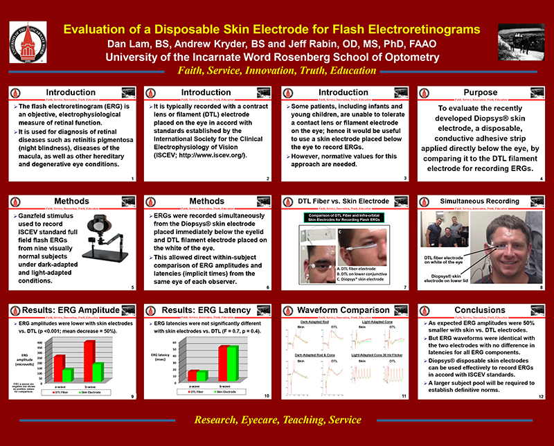
Evaluation of a Disposable Skin Electrode for Flash Electroretinograms
Conclusion: ERG amplitudes were 50% smaller with skin vs. DTL electrodes, but ERG waveforms were identical with the two electrodes with no difference in latencies for all ERG components. Diopsys disposable skin electrodes can be used effectively to record ERGs in accord with ISCEV standards.
D Lam, A Kryder, J Rabin, Presented at American Academy of Optometry
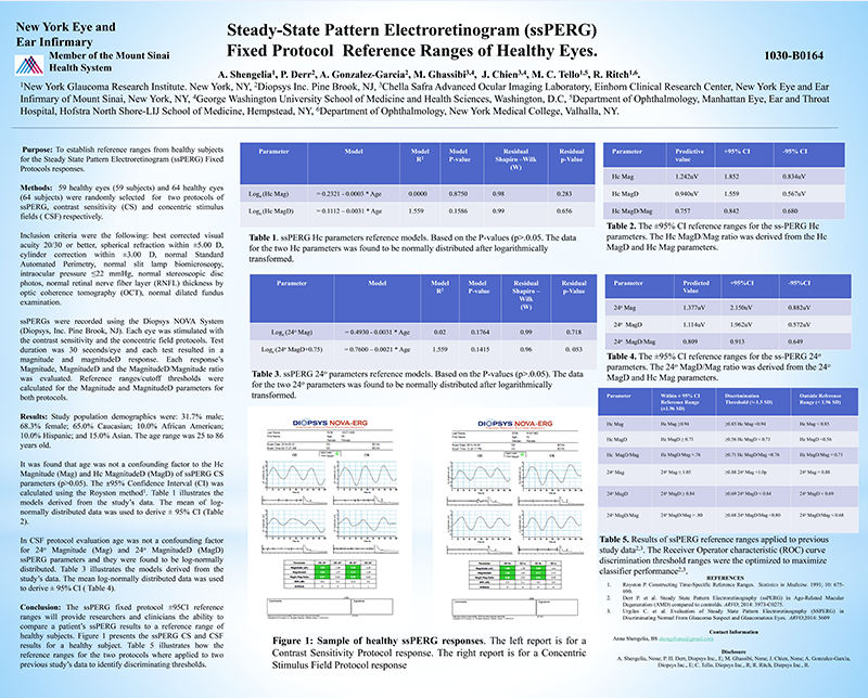
Steady-State Pattern Electroretinogram (ssPERG) Fixed Protocol Reference Ranges of Healthy Eyes
Conclusion: The ssPERG fixed protocol ±95CI reference ranges will provide researchers and clinicians the ability to compare a patient’s ssPERG results to a reference range of healthy subjects A Shengelia, P Derr, A Gonzalez-Garcia, M
Ghassibi, J Chien, M C Tello, R Ritch, Presented at the Association for Research in Vision and Ophthalmology (ARVO)
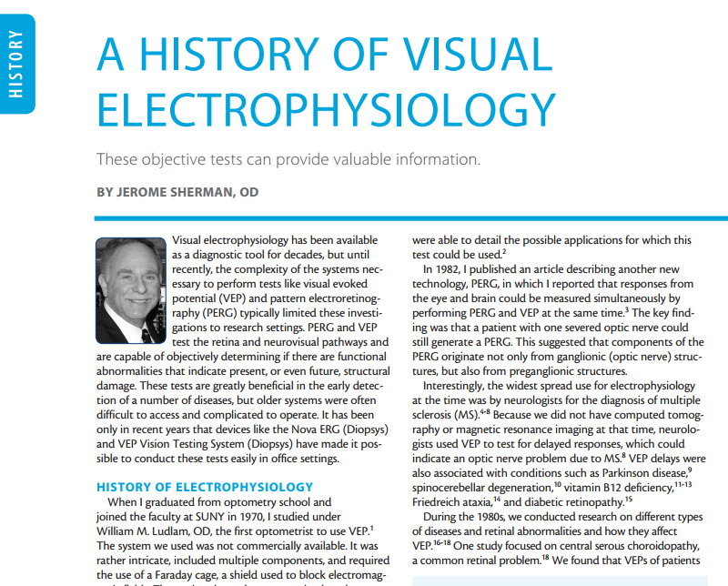
A History of Visual Electrophysiology
Jerry Sherman, OD discusses the history of visual electrophysiology, and its impact on patient care. Dr. Sherman has been using VEP since 1970, and ERG since the early ‘80s. His historical review provides background on where visual electrophysiology has been and discusses more recent advances that have made these valuable tests accessible to optometric and

Progressive Loss of Retinal Ganglion Cell Function Precedes Structural Loss by Several Years in Glaucoma Suspects
Conclusion: In patients who are glaucoma suspects, PERG signal anticipates an equivalent loss of OCT signal by several years.
M R Banitt, L M Ventura, W J Feuer, E Savatovsky, G Luna, O Shif, B Bosse, V Porciatti, IOVS

AAO Basic and Clinical Science Course on Glaucoma
Chapter 4. Section: The Glaucoma Suspect “The increasing use of short-wavelength automated perimetry (SWAP) and frequency-doubling technology (FDT) perimetry, as well as assessment of the pattern electroretinogram, may improve the ophthalmologist’s ability to recognize early glaucomatous visual function loss in patients considered to be glaucoma suspects because of a suspicious optic disc appearance.”

Electrophysiology in Glaucoma
Brian Francis, MD, discusses the use of Diopsys visual electrophysiology in glaucoma. Dr. Francis comments on the difference between transient and steady-state pattern electroretinograms (ERGs) and highlights the clinical advantages of using pattern ERG to track disease progression, detect reversal of damage in treated patients, and differentiate normal from early glaucoma.
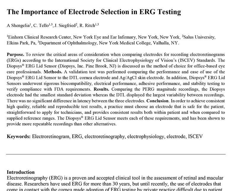
The Importance of Electrode Selection in ERG Testing
Conclusion: In order to achieve consistent high quality, reliable and reproducible test results, a practice must choose an electrode that is safe for the patient, straightforward to apply for technicians, and provides consistent results both within patient and when compared to supplied reference ranges. The Diopsys® ERG Lid Sensor meets each of these requirements, and has been shown to provide more repeatable recordings than other alternatives.
A Shengelia, C Tello, J Siegfried, R Ritch
In-office electrophysiology helps detect, monitor glaucoma damage
Brian A. Francis, MD, MS discusses how in-office pattern electroretinography helps diagnose glaucoma and track glaucomatous progression by examining retinal ganglion cells. The interview was conducted at the 2017 American Society of Cataract and Refractive Surgery meeting.
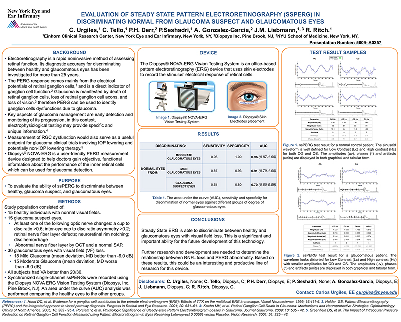
Evaluation of Steady State Pattern Electroretinography (SSPERG) in discriminating normal from glaucoma suspect and glaucomatous eyes
Conclusion: Steady State ERG is able to discriminate between healthy and glaucomatous eyes with visual field loss.
P Derr, A Gonzalez-Garcia, A Shengelia, J Chien, M Ghassibi, C Tello, R Ritch, Presented at the Association for Research in Vision and Ophthalmology (ARVO)
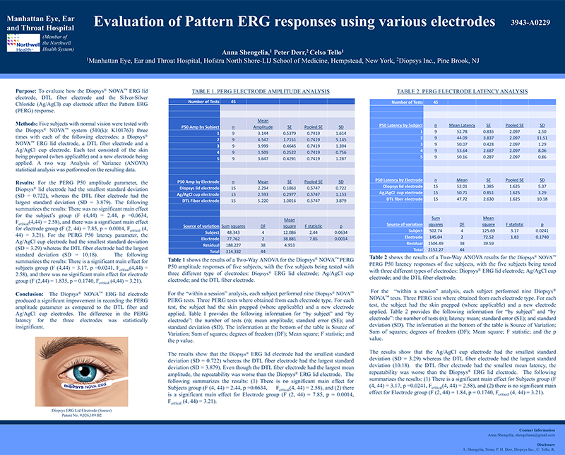
Evaluation of Pattern ERG Responses Using Various Electrodes
Conclusion: The Diopsys® NOVA™ ERG lid electrode produced a significant improvement in recording the PERG amplitude parameter as compared to the DTL fiber and Ag/AgCl cup electrodes.The difference in the PERG latency for the three electrodes was statistically insignificant.
ia, P Derr, C Tello, Presented at the Association for Research in Vision and Ophthalmology (ARVO)
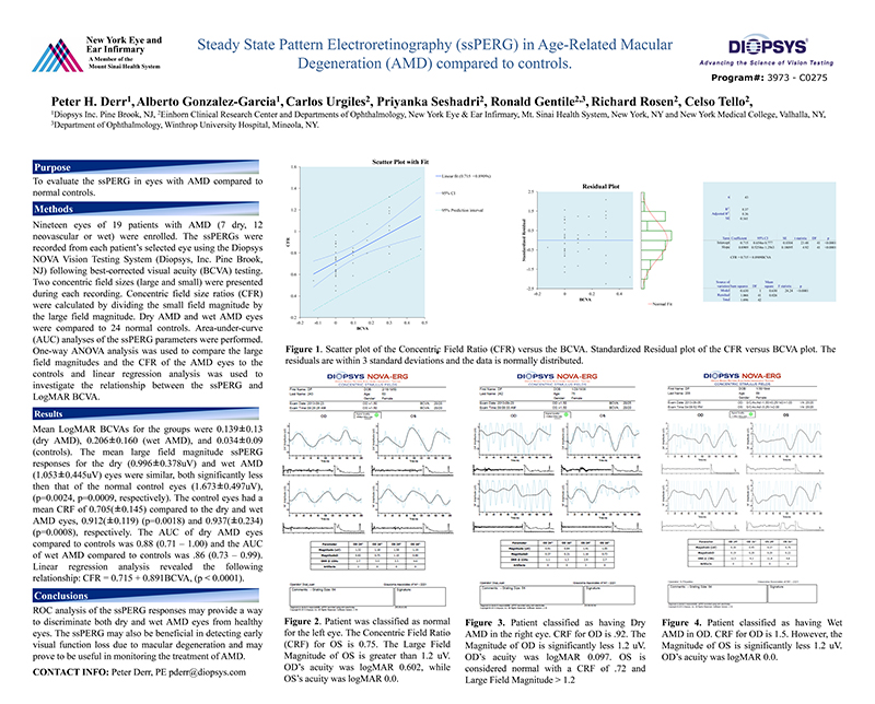
Steady State Pattern Electroretinography (ssPERG) in Age-Related Macular Degeneration (AMD) compared to controls
Conclusion: ROC analysis of the ssPERG responses may provide a way to discriminate both dry and wet AMD eyes from healthy eyes. The ssPERG may also be beneficial in detecting early visual function loss due to macular degeneration and may prove to be useful in monitoring the treatment of AMD.
P Derr, A Gonzalez-Garcia, C Urgiles, P Seshadri, R Gentile, R Rosen, C Tello, Presented at the Association for Research in Vision and Ophthalmology (ARVO)
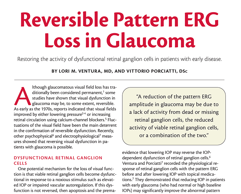
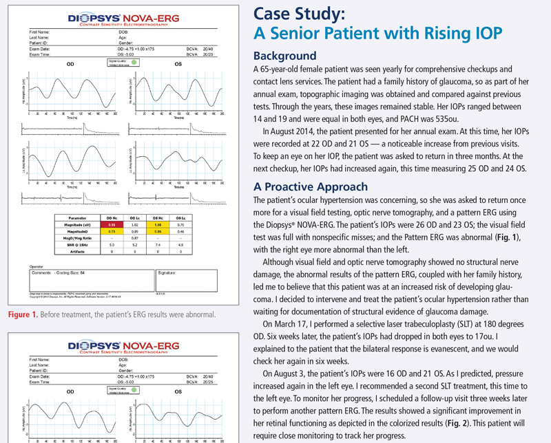
Reversible Pattern ERG Loss in Glaucoma
Clinical Implications: An improvement in pattern ERG amplitude after a reduction in IOP challenges the current opinion that structural changes of the optic nerve fiber layer precede functional changes, as measured by standard automated perimetry (SAP). It is important to consider, however, that pattern ERG and SAP probe different retinal regions and different aspects of visual function. SAP is relatively insensitive to early losses of retinal ganglion cells in the central area, where there is high retinal ganglion cell density and redundancy. In contrast, the pattern ERG, representing the summed activity of central retinal ganglion cells, may be able to discern the early generalized dysfunction of viable neurons. Given that retinal ganglion cell function is restored, at least in part, with a decrease in IOP, several multicenter studies may now have a neurophysiological basis to indicate that reducing IOP delays the onset or the progression of visual field deterioration in ocular hypertension and glaucoma.
L M Ventura, V Porciatti, Glaucoma Today
The Value of Flicker Erg B-Wave Implicit Time in Macular Degeneration
Discussion: It is well known how the presence of exudative macular degeneration in one eye is one of the major risk factors for the development of neovascularization in the fellow eye. It is thus particularly useful to have diagnostic tests that can reveal early photoreceptor alterations. Therefore, the routine use of the 30 Hz flicker in all patients with unilateral macular degeneration could be recommended for an early diagnosis of new retinal alterations.
N Pescosolido, D Rocca, D Rusciano, JSM Ophthalmology

Visually Evoked Potential Markers of Concussion History in Patients with Convergence Insufficiency
An objective visual evoked potential (VEP) marker was discovered in a high percentage of patients suffering concussion symptoms. The study authors developed a statistical algorithm from 79 patients predicting concussion history with 92 percent accuracy using the Diopsys® NOVA™ ERG and VEP Vision Testing System. These findings represent an important tool for both concussion diagnosis, and the potential for new insights and treatment options.
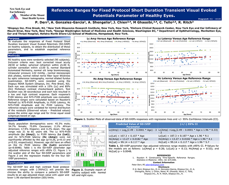
Reference Ranges for Fixed Protocol Short Duration Transient Visual Evoked Potentials Parameter of Healthy Eyes
Conclusion: The SD-tVEP low and high contrast fixed protocol reference ranges with (-/95%CI) will provide the clinician the ability to compare a patient’s SD-tVEP results to an age adjusted mean value with upper and lower 1.96 standard deviation (SD) limits.
P Derr, A Gonzalez-Garcia, A Shengelia, J Chien, M Ghassibi, C Tello, R Ritch, Presented at the Association for Research in Vision and Ophthalmology (ARVO)
Repeatability of short-duration transient visual evoked potentials in normal subjects
Conclusion: Short-duration transient VEP technology showed good within-session, inter-session repeatability, and good inter-eye correlation and agreement.
C Tello, C G V De Moraes, T Prata, P Derr, J Patel, J Siegfried, J M Liebmann, R Ritch, Documenta Ophthalmologica

Objective Assessment of Visual Attention in Mild Traumatic Brain Injury (mTBI) using Visual-Evoked Potentials (VEP)
Conclusion: VEP may be extended to assess visual attention incognitively-impaired individuals and non-verbal patients, as well as paediatric patients with attention deficit hyperactivity disorder (ADHD). Lastly, the proposed testing would also allow clinicians to evaluate objectively the effect of a visual intervention incorporating an attentional component.
N K Yadav, K J Ciuffreda, Informa Healthcare

Optimization of the Pattern Visual Evoked Potential (VEP) in the Visually-Normal and Mild Traumatic Brain Injury (mTBI) Populations
Conclusion: The use of the 20 min arc check size at both contrast levels represents an optimal clinical VEP test protocol in both the visually-normal and mTBI groups; thus, a common stimulus combination produced the largest VEP amplitude, in conjunction with normal latency values.
N Yadav, K Ciuffreda, Presented at the Association for Research in Vision and Ophthalmology (ARVO)
VEP and Human Attention: Translation from Laboratory to Clinic
Abstract: The purpose is to review recent studies that used the visual-evoked potential (VEP) to assess attention in both the visually-normal (VN) and mild traumatic brain injury (mTBI) populations. The VEP technique can be used reliably in both clinic and laboratory settings to detect attention objectively in both VN and mTBI populations.
N. K. Yadav, K. J. Ciuffreda, K. T. Willeford, P. Thiagarajan, D. P. Ludlam, Vision Development & Rehabilitation, Vol. 1, Issue 1
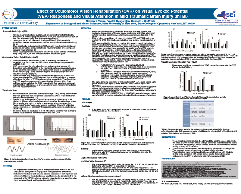
Effect of Ocular Vision Rehabilitation (OVR) on Visual Evoked Potential (VEP) Responses and Visual Attention in Mild Traumatic Brain Injury (mTBI)
Conclusion: The results demonstrate that the VEP could be used to assess objectively the effect of OVR in mTBI.
N K Yadav, P Thiagarajan, K Ciuffreda, Presented at the Association for Research in Vision and Ophthalmology (ARVO)

Effect Of Binasal Occlusion On The Visual-Evoked Potential In Mild Traumatic Brain Injury
Conclusion: There are three important clinical implications of the present VEP findings:
➢ First, an objective correlate to the decreased symptoms was found.
➢ Second, and related to the above, an objective correlate to the improved sensorimotor and visuomotor performance was found.
➢ Third, and perhaps most directly clinically relevant, in these mTBI patients with the symptom of VMS, if one does not obtain a consistent increase in VEP amplitude with BNO, the clinician should proceed with some caution; BNO may in fact be contraindicated, and this notion warrants further investigation.
N Yadav, D P Ludlam, K Ciuffreda, Presented at the Association for Research in Vision and Ophthalmology (ARVO)

Sensitivity and Specificity of Short Duration Transient Visual Evoked Potentials in Discriminating Normal from Glaucomatous Eyes
Conclusion: Short-duration transient VEP (Diopsys NOVA-LX) objectively identified decreased visual function and discriminated between healthy and glaucomatous eyes and showed good differentiation between healthy eyes and those with early visual field loss.
C Pillai, R Ritch, P Derr, A Gonzalez, L K Cox, J Siegfried, J M Liebmann, C Tello, IOVS
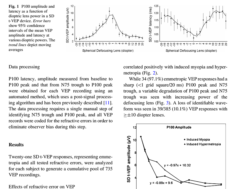
Short-duration transient visual evoked potential for objective measurement of refractive errors
Conclusion: The VEP scoring system has a potential for objective screening of RE and for a more accurate 3-step objective refraction.
A Anand, C G V De Moraes, C Teng, J Liebmann, R Ritch, C Tello, Documenta Ophthalmologica
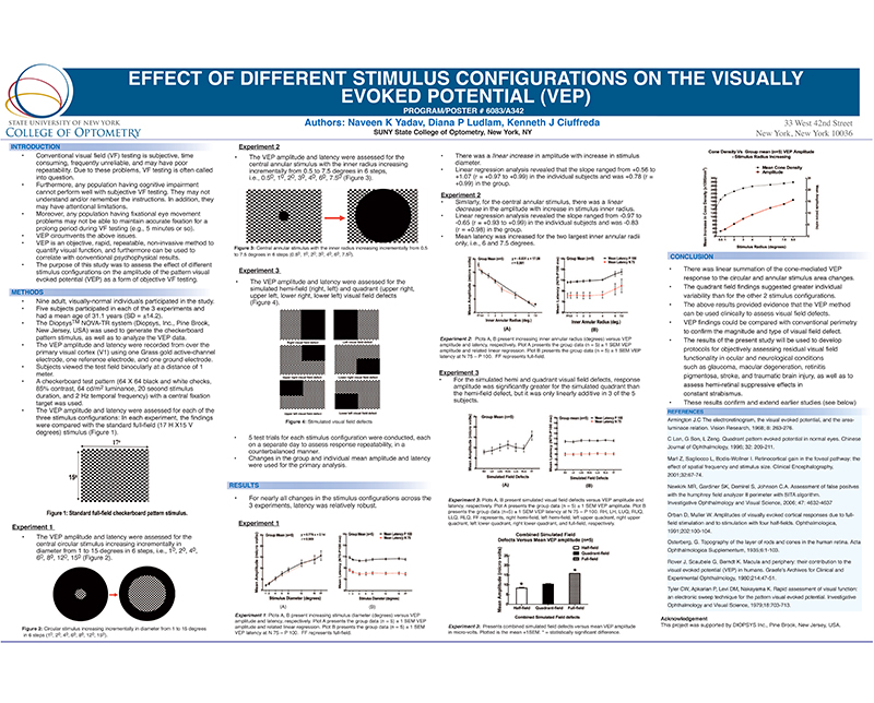
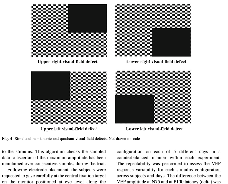
Effect of Different Stimulus Configurations on the Visually Evoked Potential (VEP)
Conclusion: The results provided evidence that the VEP method can be used clinically to assess visual field defects.
N K Yadav, D P Ludlam, K Ciuffreda, Presented at the Association for Research in Vision and Ophthalmology (ARVO)
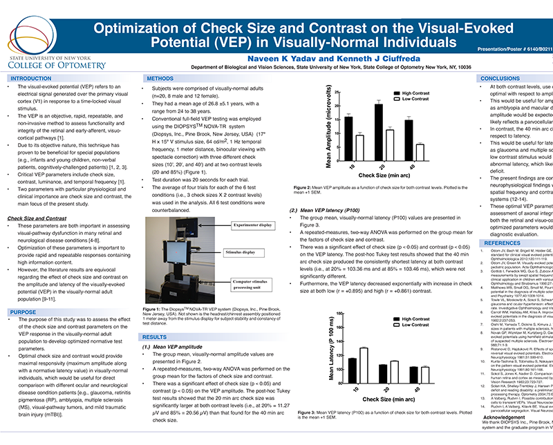
Optimization of Check Size and Contrast on the Visual-Evoked Potential (VEP) in Visually-Normal Individuals
Conclusion: Useful for amplitude-sensitive conditions, such as amblyopia and macular degeneration, and latency-sensitive conditions such as glaucoma and multiple sclerosis.
N Yadav, K Ciuffreda, Presented at the Association for Research in Vision and Ophthalmology (ARVO)
Effect of different stimulus configurations on the visual evoked potential (VEP)
Conclusion: The finding of VEP response linearity for the circular stimulus fields, and repeatability for all stimulus configurations, suggests that the clinician may be able to use the VEP technique with the suggested test patterns as a rapid and simple tool for objective assessment for several types of visual-field defects for a range of abnormal visual conditions and special populations.
N K Yadav, D P Ludlam, K J Ciuffreda, Documenta Ophthalmologica
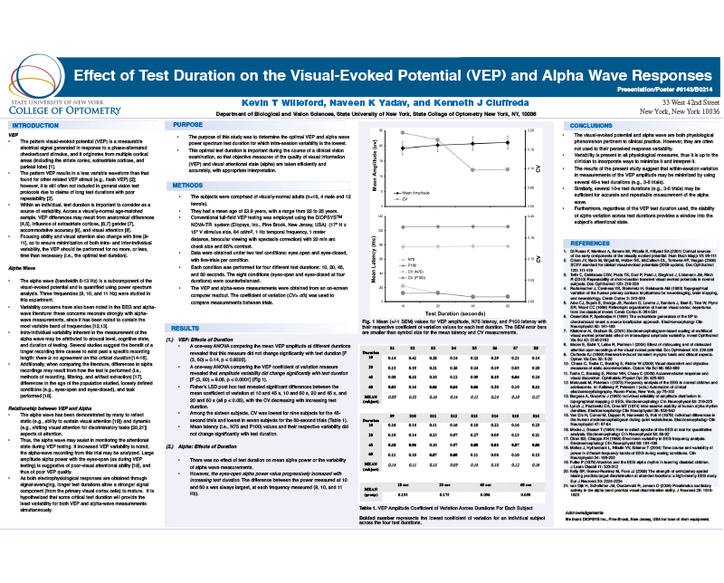
Effect of Test Duration on the Visual-Evoked Potential (VEP) and Alpha Wave Responses
Conclusion: The VEP and alpha wave are both physiological phenomenon pertinent to clinical practice. Furthermore, regardless of the VEP test duration used, the stability of alpha variation across test durations provides a window into the subject’s attentional state.
K T Willeford, N K Yadav, K J Ciuffreda, Presented at the Association for Research in Vision and Ophthalmology (ARVO)
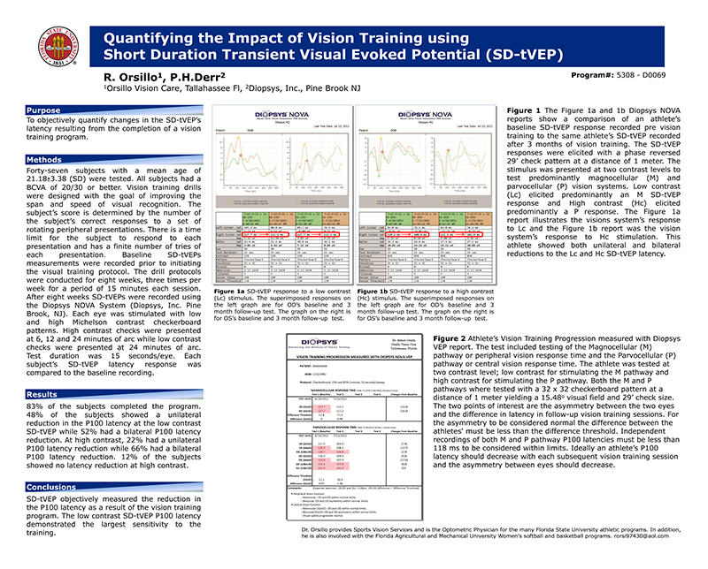
Quantifying the Impact of Vision Training using SD Transient Visual Evoked Potential
Conclusion: SD-tVEP objectively measured the reduction in the P100 latency as a result of the vision training program. The low contrast SD-tVEP P100 latency demonstrated the largest sensitivity to the training.
R Orsillo, P H Derr, Presented at the Association for Research in Vision and Ophthalmology (ARVO)
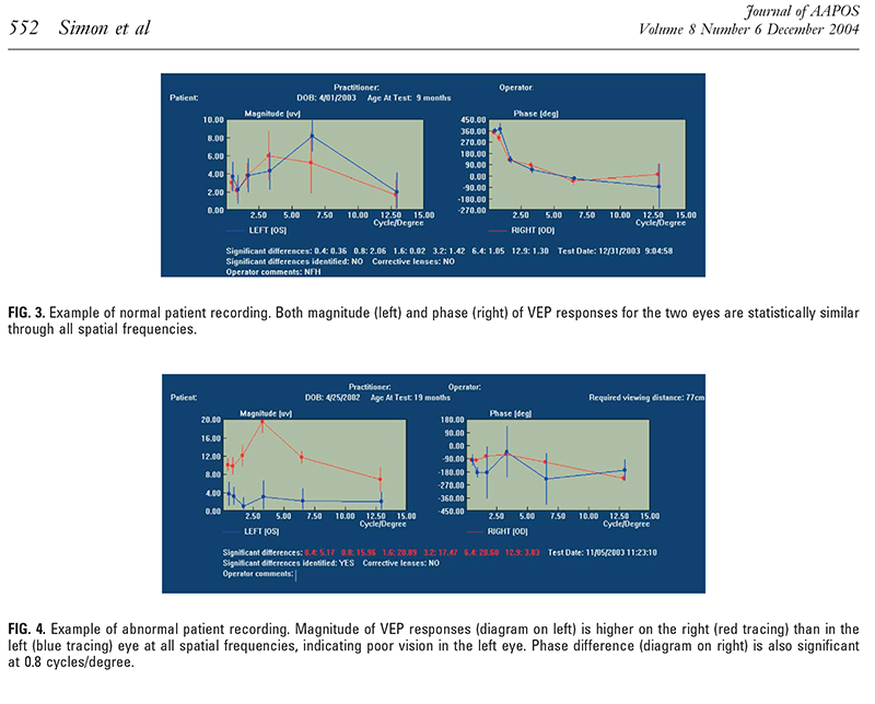
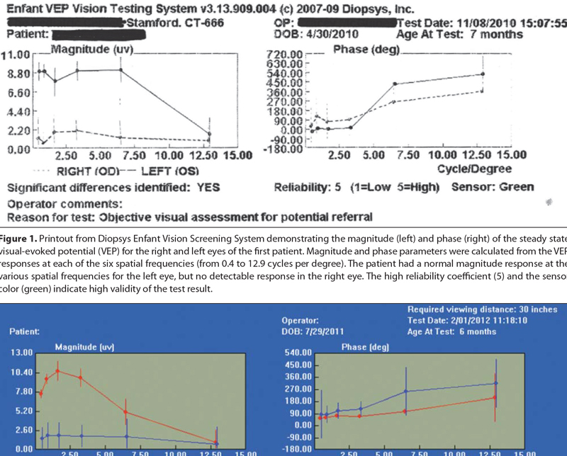
A New Visual Evoked Potential System for Visual Screening in Infants and Young Children
Conclusion: With its easy electrode placement and rapid, attractive stimulus, the new system (Enfant) overcomes technical difficulties which were associated with older VEP techniques.
J W Simon, J B Siegfried, M D Mills, J H Calhoun, J Gurland, Journal of AAPOS
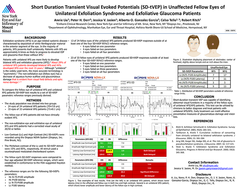
Short Duration Transient Visual Evoked Potentials (SD-tVEP) in Unaffected Fellow Eyes of Unilateral Exfoliation Syndrome and Exfoliative Glaucoma Patients
Conclusion: Short-duration transient VEP was capable of identifying abnormal visual functions in a majority of the fellow eyes of unilateral XFS/XFG patients.
A Liu, P Derr, J V Jasien, A O Gonzalez Garcia, C Tello, R Ritch, Presented at the Association for Research in Vision and Ophthalmology (ARVO)
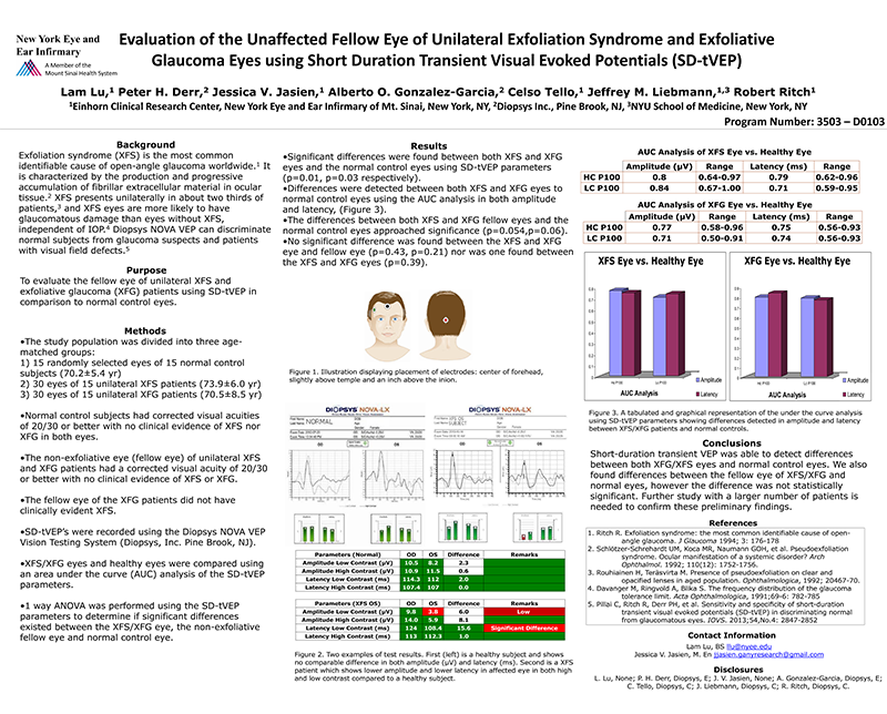
Evaluation of the Unaffected Fellow Eye of Unilateral Exfoliation Syndrome and Exfoliative Glaucoma Eyes using SD-tVEP
Conclusion: Short-Duration transient VEP was able to detect differences between both XFG/XFS eyes and normal control eyes.
L Lu, P Derr, J V Jasien, A O Gonzalez-Garcia, C Tello, J Liebmann, R Ritch, Presented at the Association for Research in Vision and Ophthalmology (ARVO)

Short Duration Transient Visual Evoked Potentials in Glaucomatous Eyes
Conclusion: In cases of asymmetric glaucoma, SD-tVEP results correlate significantly with the level of VF damage as measured by MD. In the eyes with more advanced VF loss, reduced SD-tVEP amplitude was associated with decreased macular thickness on OCT. These findings suggest that SD-tVEP may be a fast and objective method to assess or screen for functional damage in glaucomatous eyes.
T S Prata, V C Lima, C G V De Moraes, V Trubnik, P Derr, J M Liebmann, R Ritch, C Tello, Journal of Glaucoma
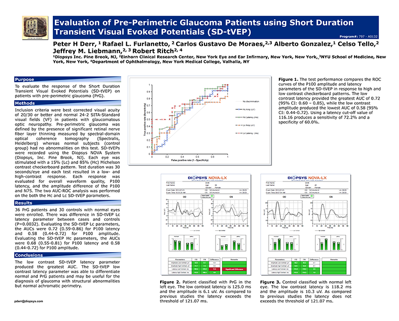
Evaluation of Pre-Perimetric Glaucoma Patients using Short Duration Transient Visual Evoked Potentials
Conclusion: The SD-tVEP low contrast latency parameter was able to differentiate normal and PrG patients and may be useful for the diagnosis of glaucoma with structural abnormalities but normal achromatic perimetry.
P Derr, F L Furlanetto, C G De Moraes, A Gonzalez, C Tello, J M Liebmann, R Ritch, Presented at the Association for Research in Vision and Ophthalmology (ARVO)
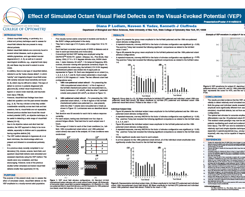
Effect of Simulated Octant Visual Field Defects on the Visual-Evoked Potential (VEP)
Conclusion: The results provided evidence that the VEP, an objective technique, could be used reliably to detect relatively small simulated scotomas in each subject. The VEP can now be extended even further and be used as a relatively rapid and simple objective technique to assess small visual-field defects
D P Ludlam, N K Yadav, K J Ciuffreda, Presented at the Association for Research in Vision and Ophthalmology (ARVO)
Sensitivity and Specificity of SD-tVEP in Discriminating Normal from Glaucomatous Eyes
Conclusion: SD-tVEP was able to identify decreased visual function objectively and to discriminate between healthy and glaucomatous eyes. In addition, it showed good differentiation between healthy and those with early field loss.
P H Derr, C Pillai, R Ritch, M Emmer, A Gonzalez, C Tello, J M Liebmann, Presented at the Association for Research in Vision and Ophthalmology (ARVO)
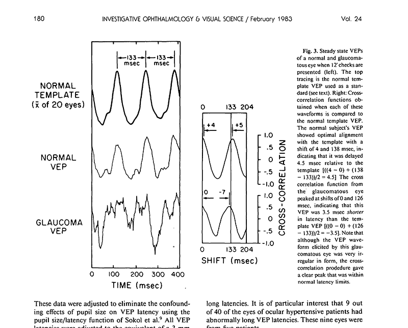
The Visual Evoked Potential in Glaucoma and Ocular Hypertension
Conclusion: The finding that is of clinical importance is the presence of abnormally long VEP latencies in some patients with ocular hypertension. The abnormal prolongation of VEP latency in these eyes may reflect subclinical optic nerve lesions that have not been uncovered with other techniques.
V L Towle, A Moskowitz, S Sokol, B Schwartz, Investigative Ophthalmology & Visual Science

Approaching the macula from the premium cataract surgeon perspective
Mitch Jackson, MD provides his perspective on facing the daily decision of whether to offer cataract surgery patients the option of multifocal IOL or other advanced IOL technology.
Mitch Jackson, MD
Healio, Ocular Surgery News, August 2016
Pattern ERG is a vital test to look at ganglion cells, says OD at the AOA Optometry’s Meeting
Nathan Lighthizer, OD, FAAO, discusses Diopsys ERG, which provides information about the inner retinal cells of the eye, as well as the Visual Evoked Potential (VEP), which measures the functional responses of the visual pathway.
Nathan Lighthizer, DD
Healio, Primary Care Optometry News, July 2016
What we “Know” is always in flux.
Paul Koch, MD shares a lesson for the ages, when evaluating glaucomatous patients. “Now I am encouraged by the use of electroretinography to objectively measure the retina’s ganglion cell layer function in patients suspected of glaucoma.”
Paul Koch, DD
Ophthalmology Management, June 2016
Early Glaucoma Detection with Pattern ERG
Robert Noecker, MD describes how pattern electroretinography can allow for earlier confirmation of glaucoma and sheds light on how information provided by pattern ERG can enable earlier intervention.
Robert Noecker, MD and Steven Vold, MD eyetube, April 2016
Using the VEP for Better Patient Outcomes in Mild Traumatic Brain Injury
An optimal VEP test protocol can differentiate objectively between visually normal patients and those with mild traumatic brain injury.
Kenneth J. Ciuffreda, OD, PhD; Naveen Yadav, BSOptom, MS, PhD; and Diana P. Ludlam, BS, COVT
Advanced Ocular Care, October 2015


Reverse-Engineering of Hyperopic Anisometropic Refractive Amblyopia
Conclusion: The VEP can facilitate decision making processes for the clinician in titrating the anisometropic prescription toward isometropia while attaining and preserving symmetry in timing and amplitude of the signals to primary visual cortex between the two eyes.
COVD, Volume 43, No. 1, 2012 – L. Press, OD, FAAO, FCOVD; D. Press, OD, FCOVD


Retinoblastoma Detected by Preschool Vision Screening Using Visual-Evoked Potentials
Abstract: Two cases of otherwise healthy children with no known family history of retinoblastoma who were diagnosed as having retinoblastoma after failing a visual-evoked potential test during a well-child visit are reported. This early detection allowed for eye-sparing treatment.
C B Estopinal, A B Wolf, S P Donahue, Journal of Pediatric Ophthalmology & Strabismus
Objective VEP Testing for Possible Malingerer Reveals Sight-Threatening Condition
This case exemplifies the necessity of an objective, functional test. The Diopsys® VEP recordings were able to uncover a serious sight and life-threatening condition in a young boy who at the time of his examination was under the care of a neurologist, reported no headaches and exhibited the embellished behaviors of a malingerer.
David Biberdorf, OD, FCOVD